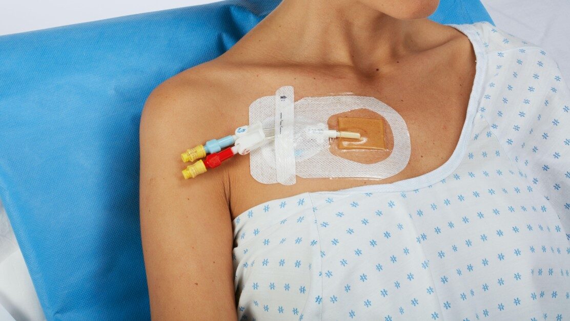
IV (intravenous) cannulation is a fundamental medical procedure used to administer fluids, medications, or blood products directly into a patient’s bloodstream.
1. Indications for IV Cannulation
✔ Fluid resuscitation (e.g., dehydration, shock)
✔ Medication administration (antibiotics, analgesics)
✔ Blood transfusions
✔ Contrast dye for imaging
✔ Continuous monitoring (e.g., ICU)
2. Equipment Needed
- Sterile IV cannula (common sizes: 18G–24G)
- Tourniquet
- Alcohol/chlorhexidine swabs
- Sterile gloves
- Transparent dressing (e.g., Tegaderm™)
- IV extension set/flush (saline or heparin lock)
3. Step-by-Step IV Insertion
A. Preparation
- Verify patient ID & consent (if applicable).
- Select vein
- Apply tourniquet (5–10 cm above site).
- Clean skin (alcohol/chlorhexidine, circular motion).
B. Cannulation Technique
- Stretch skin distal to insertion site (anchors vein).
- Insert needle (15–30° angle, bevel up).
- Look for flashback (blood in chamber).
- Advance slightly (1–2 mm further).
- Slide catheter off needle into vein.
- Release tourniquet, apply pressure proximal to tip.
- Secure with dressing, label with date & gauge.
C. Post-Insertion
- Flush with saline (check for patency & infiltration).
- Document site, gauge, and any complications.
4. Choosing the Right Cannula Size
| Gauge (G) | Color | Best For |
| 14G–16G | Orange/Grey | Trauma, rapid fluid/blood infusion |
| 18G | Green | Surgery, blood transfusions |
| 20G | Pink | Most medications, CT contrast |
| 22G | Blue | Pediatrics, fragile veins |
| 24G | Yellow | Neonates, very small veins |
5. Common Complications & Solutions
| Complication | Cause | Management |
| Infiltration | Fluid leaks into tissue | Stop infusion, elevate limb |
| Phlebitis | Vein inflammation | Warm compress, remove IV |
| Hematoma | Vein puncture/blood leakage | Apply pressure, ice |
| Occlusion | Clot in cannula | Flush gently (may need replacement) |
| Infection | Poor aseptic technique | Remove IV, antibiotics if needed |
6. Tips for Difficult IV Access
- Use warm packs (dilates veins).
- Try ultrasound guidance (for deep veins).
- Use smaller gauge (24G if fragile veins).
- Vein locator devices (infrared light helps).
Key Takeaways:
✅ Select the largest appropriate gauge for the treatment.
✅ Anchor veins properly to prevent rolling.
✅ Secure & monitor to avoid complications.
✅ Document insertion details for handover.